Andy Kin On Wong, PhDScientist, Joint Department of Medical Imaging, UHN 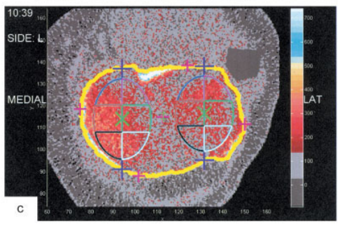 Peripheral quantitative computed tomography (pQCT) has been used to measure subchondral BMD of the knee. Although pQCT model XCT2000 has a limited gantry diameter, it can still accommodate most individuals with BMI≤ 30 kg/m2. XCT3000 has a larger gantry able to accommodate most knees even for individuals with higher BMI. Bennell previously examined subchondral BMD at the 2% and 4% tibial plateau relative to a reference line placed at a level between medial and lateral tibial compartments’ most radio-opaque plateau regions. No femoral condyles were examined, and if compartments were misaligned from the scanner’s Z-axis, the compartment-specific analyses would be oblique. https://onlinelibrary.wiley.com/doi/pdf/10.1002/art.23795 Whyte presented alternative protocols suggesting 4 individual reference lines for each of medial and lateral tibial and femoral condyles, then using 1% relative distance for tibial plateaus and 2% relative distance for femoral condyles – to account for differences in regions of interests.
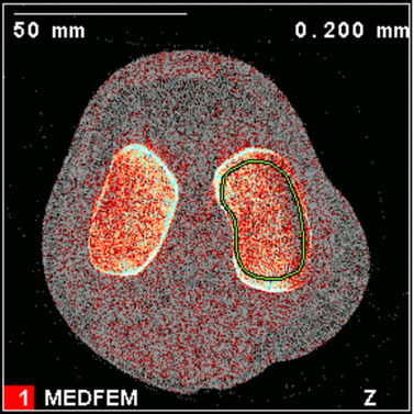
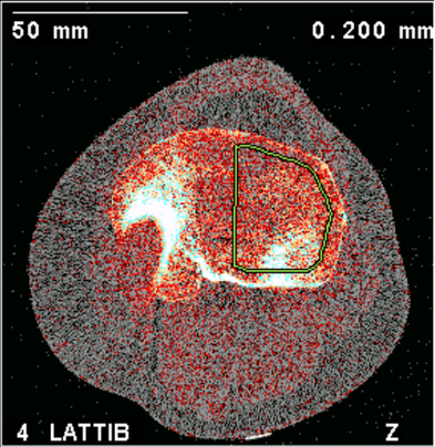
Subchondral regions of interests marked by periosteal perimeters inset by 20% and total BMD computed to yield subchondral BMD for each compartment.
The results of test-retest precision for Protocol 3 applied to healthy and diseased patients, demonstrating sufficient test-retest precision for subchondral BMD measurements overall: 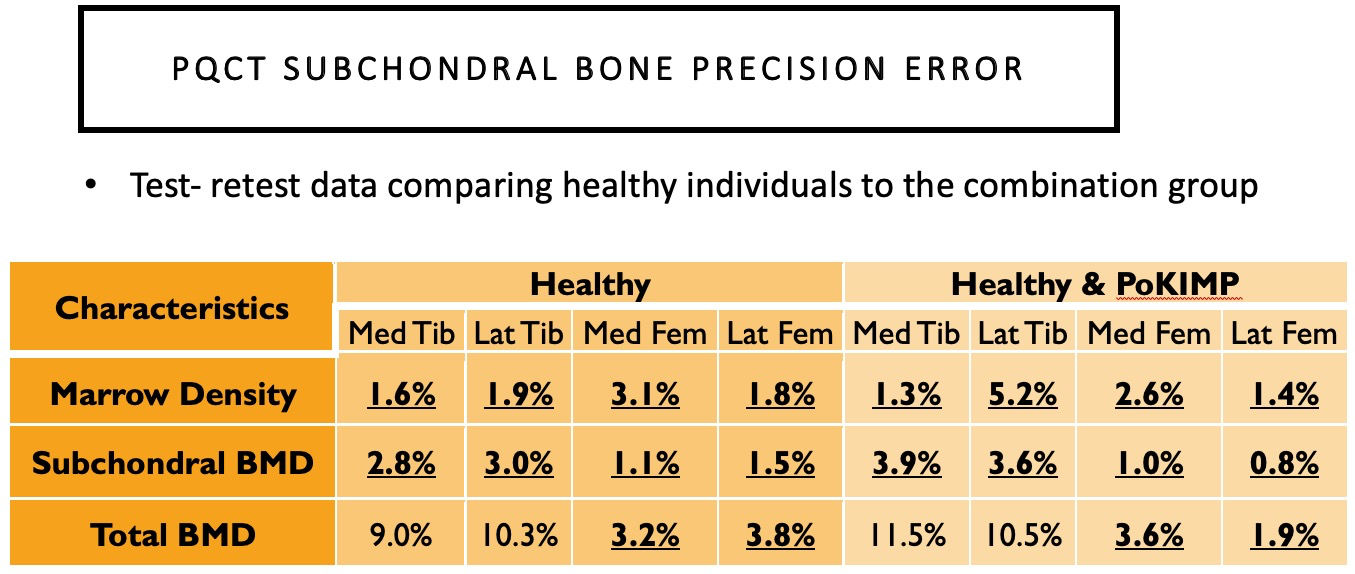
0 Comments
Leave a Reply. |
Archives
May 2024
Categories
|
||||
 RSS Feed